DNA Sequence
GAGAAGATCTGGTTTGCTGCTGCTGAGGAAGAGGAGGCTGAGGAG
DNA Output
Glu Lys Ile Trp Phe Ala Ala Ala Glu Glu Glu Glu Ala Glu Glu
Where it is produced?
DMD is a gene that stores information about a protein dystrophin. It is mainly expressed in skeletal and cardiac muscle cells, where synthesized dystrophin helps to form a dystrophin-associated protein complex. Smaller amounts of the protein are produced in the brain, where it contributes to the normal structure and function of synapses.
What's its main role?
Dystrophin is the central protein of the dystrophin-glycoprotein complex (DGC) in skeletal and heart muscle cells. Its role is to stabilize muscle cell membranes (sarcolemma) during muscle contraction and relaxation. In cardiac and skeletal muscles contraction generates mechanical force, which has to be transferred across the muscle cell without weakening or damaging the cell membrane. Dystrophin helps to achieve this by playing the role of a structural linker between the intercellular actin complex and the DGC which in turn is connected with the extracellular matrix, meaning that dystrophin in some way is a bridge between the inside of a muscle fiber and the outer supporting network. This structure allows mechanical stress and force to be spread across more evenly, reducing the risk of membrane damage. Moreover, dystrophin plays a role in membrane permeability, as when it is absent or severely reduced sarcolemma becomes fragile and more permeable, leading to abnormal leakage of minerals like calcium, which causes muscle cell damage and dysfunction.
General mechanism of action:
Dystrophin binds with actin that is inside the muscle fiber and associates with DGC in the sarcolemma, which is connected to the laminin in the outside of the cell. This creates a bridge that transmits force and organizes signalling proteins, allowing muscle fibers to stand stress and respond to mechanical force.
Functions:
- Structural Stabilization
- Force Transmission
- Molecular Scaffold
- Shock Absorber
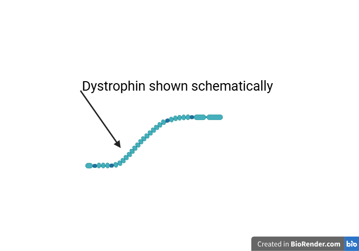
- N-Terminal Domain, that securely binds to actin filaments
- Central Rod Domain, which is a long flexible region, composed of 24 repeats and four hinge regions. These repeats act like a spring, while hinges provide flexibility
- Cysteine-Rich Domain, that is crucial for binding with sarcolemma
- C-Terminal Domain interacts with dystrobrevin and syntrophins, strengthening the complex overall
- Duchenne muscular dystrophy (DMD)
- Becker muscular dystrophy (BMD)
- DMD-associated dilated cardiomyopathy (DCM)
Structure:
Dystrophin is made from four primary domains:
Related diseases:
The dystrophinopathies cover a spectrum of X-linked muscle disease that ranges from mild to severe and includes Duchenne muscular dystrophy (DMD), Becker muscular dystrophy (BMD), and DMD-associated dilated cardiomyopathy (DCM)
DMD is caused by mutations in the DMD gene that result in an almost complete absence of functional dystrophin. Without the protein the muscle cell membrane is easily damaged during contraction, leading to muscle fiber injury and replacement of muscle tissue by fat and fibrosis. This causes severe muscle weakness that usually begins in early childhood, with symptoms such as frequent falls, difficulty in running and later loss of walking ability. Over time the disease also affects the heart and respiratory muscles, leading to breathing difficulties and reduced life expectancy.
BMD is also caused by mutations in the DMD gene, but in this case some dystrophin is still produced. And so as dystrophin still functions but in less amounts, muscle damage occurs more slowly. Symptoms usually appear later and include muscle weakness, cramps, and difficulty with activities. However, even with skeletal muscle symptoms being relatively moderate serious heart problems such as dilated cardiomyopathy can still develop.
In DMD-associated DCM, the dystrophin defect mainly impairs the function of cardiac muscle cells. Because dystrophin is abnormal, the membranes of heart muscle cells are less stable during repeated contraction, which leads to ongoing damage of the heart chambers and reduced efficiency of pumping. The main symptoms are related to heart dysfunction and may include fatigue, shortness of breath, reduced exercise tolerance, arrhythmias, and swelling. The major consequence is progressive weakening of the heart, which can become life-threatening.